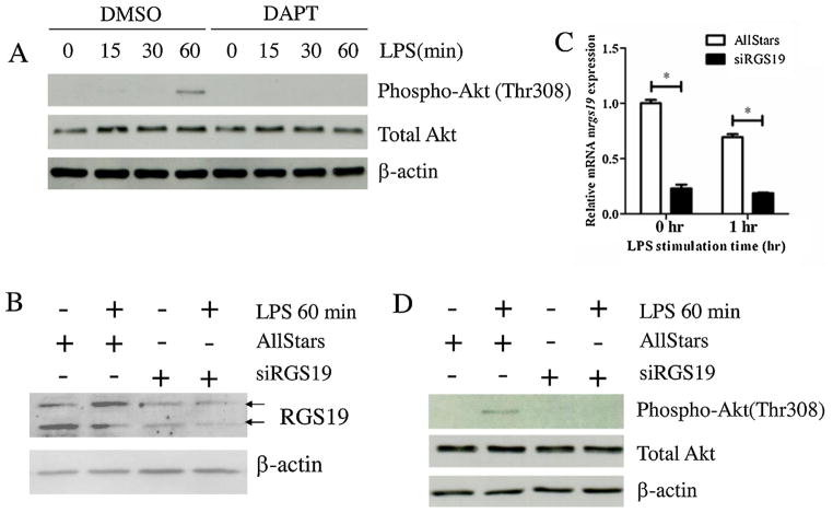
Fig. 3.
The Effect of DAPT and silencing of RGS19 on Akt phosphorylation in LPS-stimulated macrophages. (A) The RAW264.7 cell line was pretreated with DAPT (25 μM) or the vehicle control (DMSO) for 1 h before the cells were stimulated with LPS (100 ng/ml) for the indicated times. Phosphorylation of Akt in cell lysates was detected using Western blotting. (B) RAW264.7 cells were transfected with the control scrambled siRNA or rgs19-specific siRNA. After the transfection, cells were stimulated with 100 ng/ml LPS for 0 and 60 min, and protein lysates were collected to examine the level of RGS19 expression using Western blotting. Arrow heads indicated the two bands corresponding to phosphor-RGS19 and total RGS19. (C) RAW264.7 cell line was transfected with siRNA, as described above, and stimulated with LPS (100 ng/ml) for 0 and 60 min. Total RNA was harvested to investigate the expression level of rgs19 mRNA by qPCR. * indicates statistical significance (p < 0.05). (D) RAW264.7 cell were transfected with siRNA, as described above, and stimulated with LPS (100 ng/ml) for 0, 15, 30 and 60 min. Phosphorylated (Thr308) and total Akt were detected using Western blotting.

