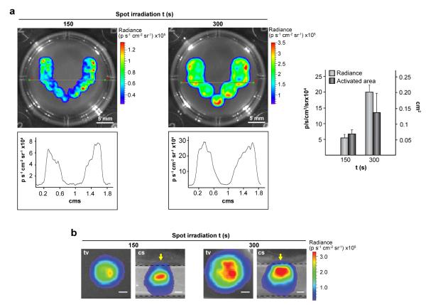
Fig. 3.
Patterning transgene expression using optical hyperthermia. C3H/T101/2-fLuc cells were encapsulated within hydrogels containing 0.03 mg mL−1 HGNPs and cultured for 24 h. Constructs were treated with rapamycin, NIR-irradiated at 44 mW mm−2 at each spot for the indicated times and cultured further for 24 h before bioluminescence imaging. (a) Top view of hydrogels NIR-irradiated at 11 (left) or 7 (right) adjacent spots (upper panel), and quantification of fLuc activity in the construct midline (lower panel). Graph shows the average luminescence radiance levels and the size of activated areas in induced regions. Each value represents the mean ± SD. (b) Top (tv) and cross-section (cs) views of a single spot exposed to NIR irradiation. Arrows in cs views indicate the direction of NIR laser incidence. Dashed lines delimit the edges of the hydrogel. Scale bars = 1 mm.

