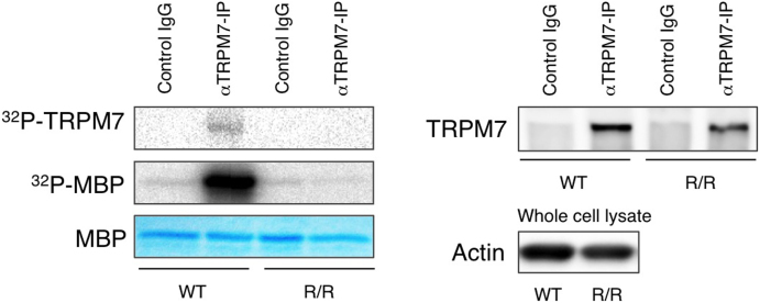
Figure 3. TRPM7 kinase activity in TRPM7R/R mice.
Kinase assay of TRPM7 proteins from WT and TRPMR/R mouse embryonic fibroblasts (MEF). Left panel shows 32P incorporation into autologous TRPM7 and exogenous myelin basic protein (MBP) by TRPM7 immunoprecipitated from WT or TRPM7R/R MEF lysates. Coomassie blue staining of MBP was used to ensure equal quantities of MBP. Right panel shows Western blot analysis of TRPM7 levels in the immunoprecipitates used in the left panel. Probing for actin in the whole cell lysates was used to ensure the presence of equal amount of protein in the lysates before immunoprecipitation. Full length blots and gel images are shown in Fig. S4.

