Figure 2.
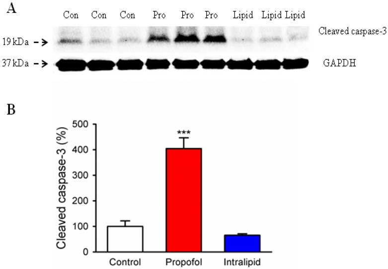
Prenatal propofol exposure induced caspase-3 activation in the brain tissues of fetal rats. A 2 h IV infusion of Propofol (Pro), intralipid (Lipid) or saline control (Con) was administered to pregnant rats on G18 (N = 3 dams per group). Fetuses were removed via C-section at six hours post infusion. Whole cerebral hemispheres of fetal rats were harvested and analyzed by Western blot. (A) Propofol infusion induced significantly higher caspase-3 activation in the brain tissues of fetal rats as compared to the control condition or intralipid infusion with Western blot analysis. There is no statistically significant difference in the amounts of GAPDH in the rat brain tissues following the propofol, intralipid treatment or control condition. (B) Quantification of the Western blot shows that propofol anesthesia increased cleaved caspase-3 levels in the rat brain tissues compared to the control groups (*** p < 0.001). Intralipid infusion did not affect the levels of cleaved caspase-3 in the brain of fetal rats. Data from six pups (2 pups from 3 litters) per group are expressed as means ± SEM.

