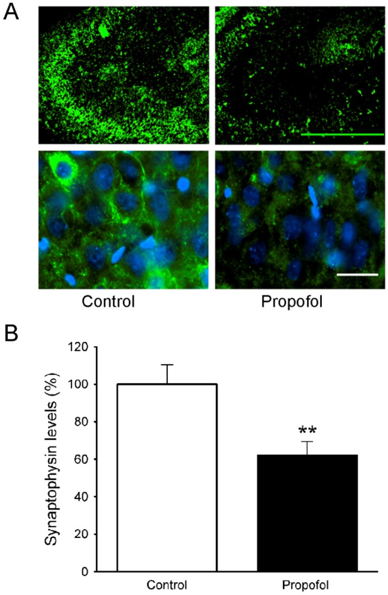
Figure 5.
Propofol administered to pregnant rats decreased synaptophysin levels in the hippocampus of the offspring rats. (A) Immunofluorescent staining for synaptophysin in the brain tissues of offspring rats on P28. Left panel: control; right panel: propofol. Scale bar = 100 µm. (B) Semiquantitative analysis of the immunohistochemistry image shows that the synaptophysin levels in the brain of rats exposed to propofol in utero are significantly lower than that of control rats (** p = 0.009 significant difference from age-matched control group; N = 6 litters, 1 offspring per litter, n = 6 per group).

