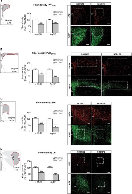
Figure 4. Maternal HFD-feeding exclusively during lactation impairs axonal projections of ARH neurons to intrahypothalamic target sites.
Images and quantification of α-melanocyte-stimulating hormone (α-MSH) and agouti-related peptide (AgRP) immunoreactive fibers innervating (A) the anterior endocrine paraventricular nucleus of the hypothalamus (PVHant; nα-MSH =6vs7 and nAgRP=7vs7), (B) the posterior preautonomic PVHpost (nα-MSH =5vs5 and nAgRP=4vs4), (C) the dorsomedial nucleus of the hypothalamus (DMH; nα-MSH =7vs7 and nAgRP=4vs5) and (D) the lateral hypothalamic area (LH; nα-MSH =6vs6 and nAgRP=6vs4) at 8 weeks of age. Schematics illustrating the localization in the CNS of the respective hypothalamic nuclei presented in the pictures were based on and modified from Brain Maps: Structure of the Rat Brain (Swanson, 1998). Coordinates were adapted according to the Mouse Brain in Stereotaxic Coordinates (Franklin and Paxinos, 1997). White boxes indicate area of quantification. 3V = third ventricle, fx = fornix. Scale bar = 100 μm. Data are presented as mean ± SEM, *p < 0.05. **p < 0.01 versus all other groups of offspring.

