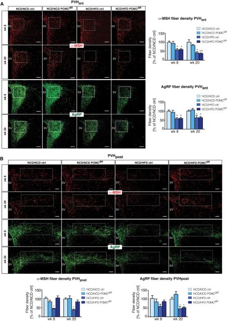
Figure 6. POMC-specific IR-deficiency in NCD/HFD offspring rescues POMC axonal projections to preautonomic regions in the PVH.
Images and quantification of α-melanocyte-stimulating hormone (α-MSH) and agouti-related-peptide (AgRP) immunoreactive fibers innervating (A) the anterior neuroendocrine paraventricular nucleus of the hypothalamus (PVHant) at 8 (nα-MSH and nAgRP =8vs8vs8vs10) and 20 weeks of age (n = 5 for all groups); and (B) the posterior preautonomic PVH (PVHpost) at 8 (nα-MSH =7vs6vs5vs6 and nAgRP=5vs6vs5vs6) and 20 weeks of age (n= 5 for all groups). White boxes indicate area of quantification. 3V= third ventricle. Scale bar = 100 μm. Data are presented as mean ± SEM, *p < 0.05 versus all other groups of offspring, unless otherwise indicated. See also S5 for images and quantification of α-MSH and AgRP immunoreactive fibers innervating the dorsomedial nucleus of the hypothalamus (DMH) and the lateral hypothalamic area (LH) at 20 weeks of age.

