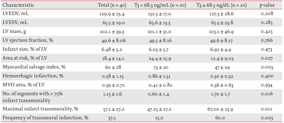
Table 3.
Results of cine-magnetic resonance imaging (MRI), T2-weighted MRI, and contrast-enhanced MRI, in terms of T3 level

Values are presented as mean ± SD.
T3, triiodothyronine; LVEDV, left ventricular end-diastolic volume; LVESV, left ventricular end-systolic volume; LV, left ventricle; MVO, microvascular obstruction.
