Fig. 7.
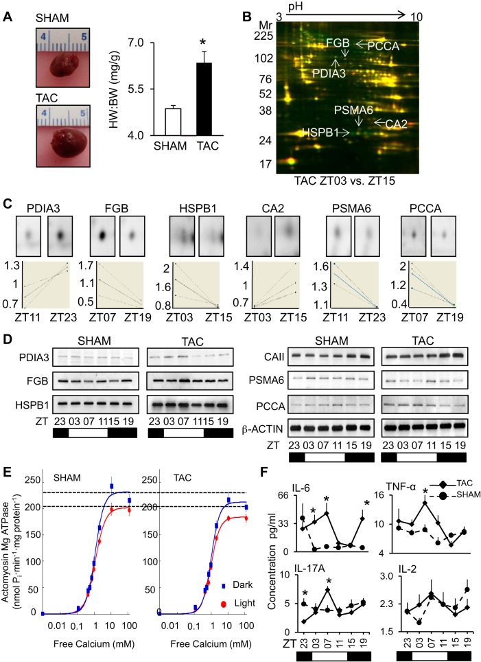
Altered day/night protein profiles in TAC heart. To demonstrate a time-of-day proteome underlying heart disease, we used a model of compensated heart disease (transverse aortic constriction; TAC). A: representative hearts from mice at 1 wk post-TAC were enlarged (left, top) vs. sham (left, bottom), and characterized by cardiac hypertrophy (right). TAC, black, sham, white. *P < 0.05; n = 36 (18 TAC, 18 sham). HW, heart weight, mg; BW, body weight, g. See Supplemental Data S6 for pathophysiology data (morphometrics, echocardiography, and hemodynamics). B: proteins with altered abundance in hearts at 1 wk post-TAC at different time points across the diurnal cycle were identified by 2D-DIGE. and C: DeCyder bioinformatics (top, single-channel spot images; bottom, graph views). D: independent validation of DeCyder bioinformatics by Western blot, using β-ACTIN as a loading control. One-week TAC profiles were further compared with the sham profiles on Western blots. See Supplemental Data S1 and S3 for tryptic peptide hits from LC/MS/MS, protein identification, and DeCyder data. E: hearts at 1 wk post-TAC were maintained with diurnal variation in myofilament activity, as measured by actomyosin MgATPase, but the diurnal calcium profiles were attenuated; also see Supplemental Data S7. F: compared with shams, the 1 wk post-TAC mice had altered diurnal profiles in serum cytokines important for early remodeling, as measured by multiplex ELISA (also see Supplemental Data S8). Values are expressed as means ± SE. *P < 0.05; n = 6 samples per time point, 6 time points total.

