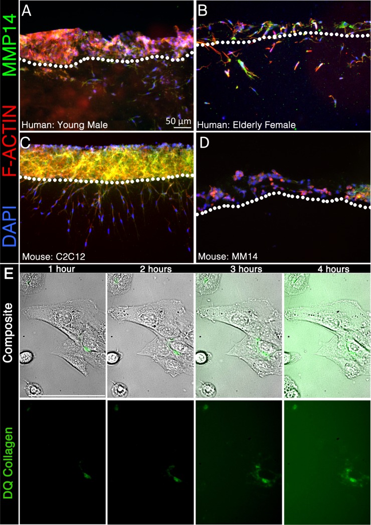
Fig. 4.
All human satellite cell populations tested express MMP-14 and invade collagen I, while murine cell lines differ from primary cells and each other. Two primary (nonclonal, nontransformed) human myoblast samples from a 5-yr, 5-day-old male (A) and a 73-yr-old female (B) and two immortalized murine myoblast cell lines, C2C12 (C) and MM14 (D), were seeded on a 3D collagen type I matrix for 90 h and stained for MMP-14 (green) with phalloidin 635 (red) and DAPI (blue). E: images are reference stills from Supplemental Video S1. The panel illustrates collagen proteolysis (green) by C2C12 myoblasts over 4 h. A composite (top) of the FITC (bottom) and brightfield channels shows localized proteolysis respective to the cell. C2C12 satellite cells adhered to the bottom of a 48-well plate were challenged with 3D collagen I copolymerized with DQ Collagen I (1 mg/ml; green.)

