
Fig. 2.
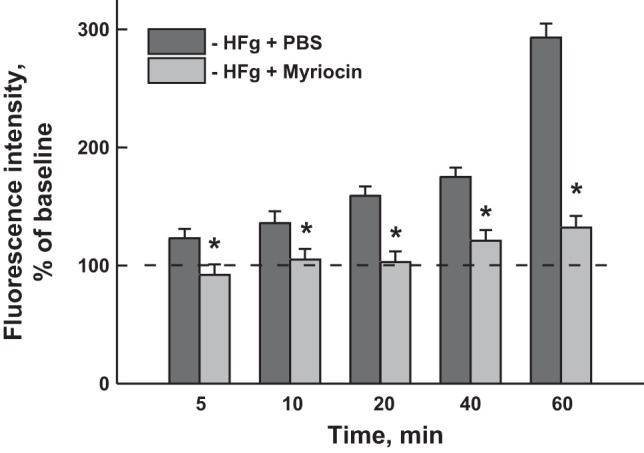
Cerebrovascular permeability to macromolecules in hyperfibrinogenic (HFg) mice. Pial venular permeability to FITC-BSA was assessed in HFg mice treated with myriocin (0.5 mg·kg−1·day−1) or PBS for 3 days. Fluorescence intensity changes in an area of interest adjacent to the venular segment were measured as described in methods. Venular permeability was assessed by changes in the ratio of fluorescence intensity measured in the interstitium adjacent to the venule to that inside the vessel. Values (means ± SE) are shown as percent change in fluorescence compared with PBS alone (control); n = 4. *P < 0.05 vs. HFg + PBS.