
Fig. 10.
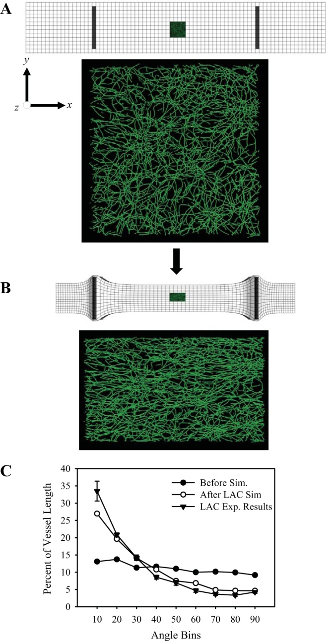
Anisotropic deformation of the matrix due to cellular contractility is sufficient to cause microvascular alignment, independent of any other cellular behavior. Mapping the displacement field from the LAC FE model onto a distribution of randomly oriented microvessels results in a new alignment pattern that closely resembles alignment within the experimental LAC constructs. A: day 0, initial mesh geometry with the microvessel dataset positioned at geometric center. B: day 6, with the use of FE displacement field to map the microvessels, a new alignment pattern emerged due to contraction in the Y- and Z-directions. C: length-normalized distributions microvessel angle with the long axis (x-axis). The 10° angle bin represents angles from 0° to 10°, which were parallel to the long axis (x-axis). The 90° angle bin represents angles from 80° to 90° and was parallel to the short axis (y-axis). The angle orientation distribution before mapping (●) indicates random microvessel orientation. After mapping (○), microvessels were preferentially aligned along the long axis. Orientation data from LAC construct experiments are shown as well (▲). Error bars indicate standard error. Sim, simulation.