
Fig. 5.
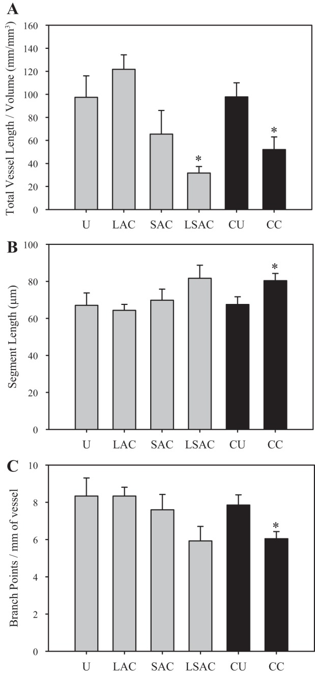
Quantitative measurements of microvessel growth for different boundary conditions. A: microvessel vascularity tended to decrease as gels became more constrained. Vascularity for the long-short axis constrained (LSAC) group was significantly lower than that in the U (*P = 0.004) and LAC groups (*P = 0.044). Vascularity in the CC boundary condition was significantly less than that in the CU condition (*P = 0.017). B: average segment length tended to increase as the gel became more constrained. Segment length in the CC cultures was significantly greater than that in the CU cultures (*P = 0.043). C: microvessel branching tended to decrease as the gel became more constrained. Branching within the CC boundary condition culture was significantly reduced relative to the unconstrained control, CU (*P = 0.020). Bars indicate standard error.