
Fig. 8.
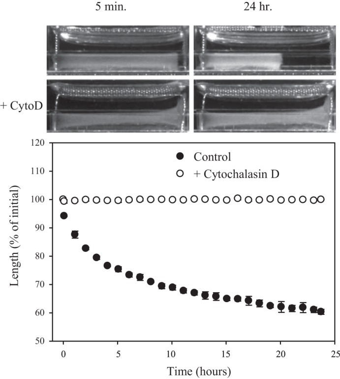
Contraction of LAC vascularized gels after cutting their attachment to the boundary. To determine whether LAC vascularized gels were under elastic stress due to cell-induced compaction, gel cutting experiments were performed. After 4 days in vitro, LAC gels in the control group were given fresh media, whereas LAC gels in the Cytochalasin D (CytoD) group received fresh media plus 10 μM CytoD. CytoD inhibits actin polymerization. Twenty minutes later, 1 end of the LAC gels was cut free from the boundary constraint using a scalpel. Gels were returned to the incubator, and digital images were captured 5 min later. Imaging continued every 20 min (72 images over 24 h). A glass mirror under the gels was used to obtain multiple culture dimensions. Culture length, height, and width were measured with ImageJ calibrated to optical markers in the same focal plane. Top: control LAC gel at 5 min and 24 h after separation from the boundary constraint. Middle: LAC gel treated with CytoD at 5 min and 24 h after separation. Bottom: remaining length (% of initial length) versus time after cutting. Neither the control nor the CytoD-treated gels exhibited any significant contraction at 5 min after separation. Over the subsequent 24 h, control LAC gels contracted 40% along the long axis. In contrast, the CytoD-treated LAC gels did not exhibit any measureable contraction. These results verify that little if any elastic stress accumulates within the extracellular matrix (ECM) of LAC gels during the culture period, and contraction after separation from the boundary constraint is due to cell contractility.