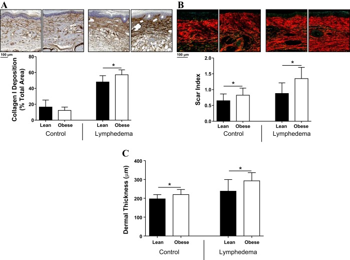
Fig. 4.
Obese mice have increased fibrosis after lymphatic injury. A: representative photomicrographs (×20 magnification) and quantification of collagen type I deposition in control and lymphedematous animals 6 wk postoperatively. *P < 0.05. B: representative photomicrographs (×20 magnification) and quantification of scar index (Sirius red staining) in control and lymphedematous animals 6 wk postoperatively. *P < 0.05. C: quantification of dermal thickness in control and lymphedematous animals 6 wk postoperatively. *P < 0.05.

