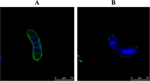
Figure 2.

Immunofluorescent detection of native Ts-Pmy expressed on the surface of T. spiralis ML using the anti-Ts -Pmy C9 binding domain mAb 9G3. The longitudinal section of ML was incubated with mAb 9G3 (5 μg/ml), and subsequently incubated with Alexa Fluor 488-labeled goat anti-mouse IgG antibody (in green) or DAPI to label nuclei (in blue) (A). Normal mouse serum at the same dilution was used as a negative control (B). The scale bars represent 15 μm.
