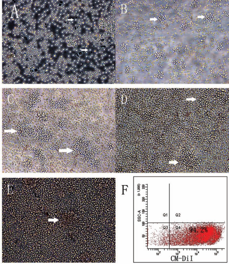
Fig. (1).
The morphology of adult hPBMCs in pre-induction culture. (A) Freshly isolated adult hPBMCs after seeding and exposure to the conditioned neural stem cell medium (x 200 Magnification). (B-E) Colonies (cell clusters, white arrows) increased in number and size overtime. (B) 24 h, (C)48 h, (D)three days, and (E) four days in conditioned medium (x 200 Magnification). (F) The labeling rate by CM-DiI as measured by flow cytometry.

