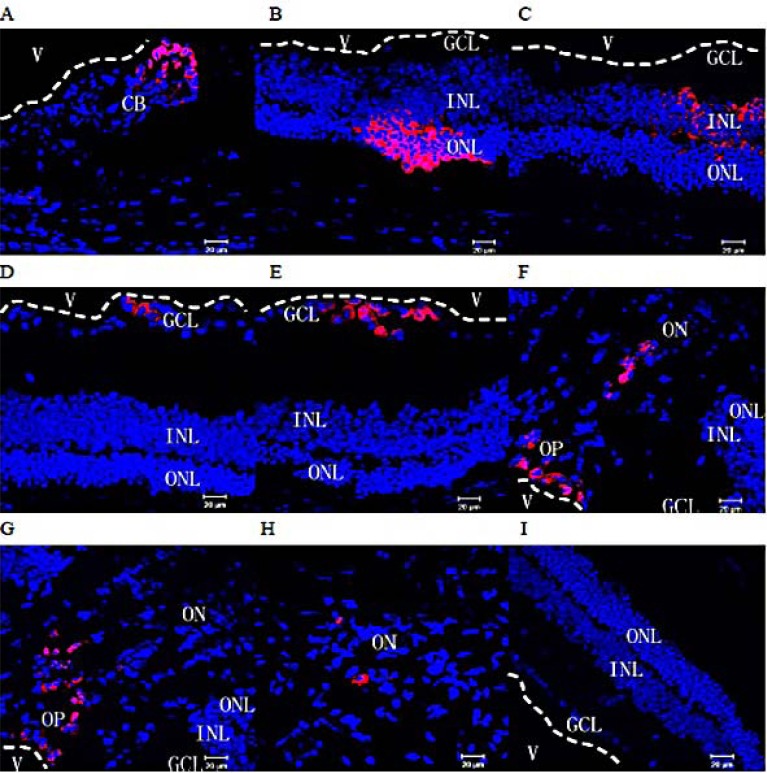
Fig. (3).
Migration within the eyeball of rds mice. Two and three months after subretinal transplantation into rds mice, the distribution of transplanted cells labeled with CM-Dil (red) was assessed in frozen retinal sections. Many transplanted cells were observed in aggregates in the ciliary body (A), the retinal outer nuclear layer (B), inner nuclear layer (C), ganglion cell layer (D&E), optic papilla (F&G), and within the optic nerve (H). (I) No CM-DiI-labeled cells were found in the retinas of rds mice from the control group (red: CM-Dil-labeled cells; Blue: cell nuclei counterstained with Hoechst 33342; V: vitreous cavity. The white dotted lines mark the boundaries of the vitreous cavity and the retina; CB: ciliary body; ONL: outer nuclear layer; INL: inner nuclear layer, GCL: ganglion cell layer, OP: optic papilla, ON: optic nerve). All scale bars represent 20 μm.

