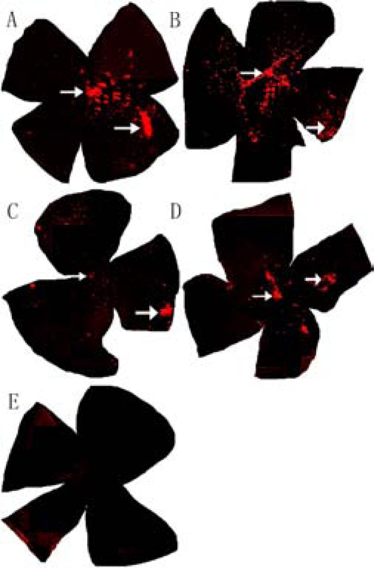
Fig. (4).
The extent of radial migration within retinas of rds mice. Two and three months after subretinal transplantation, the distribution of CM-DiI-labeled transplanted cells (red) was assessed in whole-mount retinas from the treated group (× 50 Magnification). Most of the transplanted cells remained near the injection site (white arrow in the right section of each specimen) but aggregates were also found in the optic papilla (white arrows at the center). CM-DiI staining was more intense and widespread after 2 months (A&B) than after 3 months (C& D). (E) There were no CM-DiI-labeled cells in whole-mount retinas from control group rds mice.

