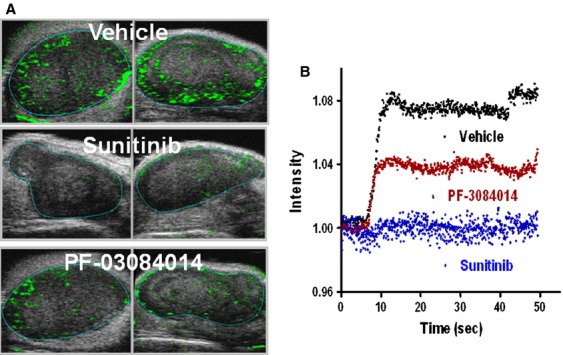
Figure 5.

Contrast-enhanced ultrasound imaging to detect the functional vasculature changes in PF-03084014 or sunitinib treated MDA-MB-231Luc tumor. Mice bearing tumors in the range between 250–400 mm3 treated until day 4. Mice were i.v. injected with 100 μL micro-bubble solution prior to imaging. N = 6 mice/group. (A) Representative images of each group. (B). Time-intensity curves depict the blood vessel perfusion rate change after the microbubble injection.
