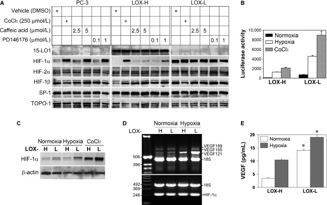
Figure 1.
Stable 15-LO1 transfection altered HIF-1α and HIF-1 transcriptional activity. (A) Western blotting analysis of crude nuclear extracts from PC-3, LOX-H and LOX-L cells. The cells in six-well plates at 70% confluence were subjected to overnight serum starvation, and treated with different reagents in serum-free media for 16 h before harvest. 15-LO1 inhibitors Caffeic acid and PD146176 were dissolved in dimethyl sulfoxide (DMSO). DMSO volumes were <0.5% (v/v) in culture medium. Immunoblots were repeatedly stripped and probed. (B) Transient transfection and reporter gene assays were conducted in LOX-H and LOX-L cells. After transfection for 24 h, cells were cultured for additional 16 h under normoxic or hypoxic conditions, or treated with CoCl2, before harvest. The figure represents mean ± SD of triplicate of one experiment. (C) Whole cell lysates in the transient transfection assay in B were analyzed for HIF-1α expression by Western blot. Similar results were shown in triplicate of one experiment, and reproduced in two more repeats. (D) Total RNA in LOX-H and LOX-L cells was analyzed for transcription of the VEGF (upper panel) and HIF-1α (lower panel) gene by RT-PCR after cells were subjected to normoxia or hypoxia for 24 h. DNA standards and the products of major VEGF isoforms are indicated. (E) Culture medium in experiment D was assayed for VEGF production with ELISA. Bars indicate standard deviations of triplicates and asterisk (*) denotes statistical significance compared to those in LOX-H cells (P < 0.05).

