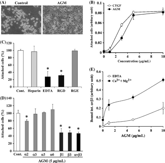
Figure 4.

Effects of AGM on adhesion of human vascular endothelial cells. (A) HUVECs suspended in serum-free basal medium were incubated on plates precoated with PBS alone (Control) or 10 μg/mL AGM, and their phase-contrast micrographs were taken after for 2-h incubation. Original magnification, ×300. (B) Ninty-six-well ELISA plates were precoated with the indicated concentrations of AGM (closed circles) or CTGF (open circles). HUVECs were incubated for 2 h on the coated plates. The relative number of adherent cells was measured as described in Materials and Methods. Each point represents the mean ± SD of the numbers of cells in triplicate wells. (C) Effects of heparin, EDTA, and RGD/RGE peptides on attachment of HUVECs to AGM. HUVECs were incubated for 2 h in the absence (Control) or presence of 100 μg/mL heparin, 10 mmol/L EDTA, 1 mmol/L RGD, or 1 mmol/L RGE on the plates precoated with 5 μg/mL AGM. The cells attached to the plates were measured as described above. *P < 0.01. (D) Effects of function-blocking anti-integrin antibodies on cell adhesion to AGM. HUVECs suspended in serum-free basal medium were incubated with control mouse IgG (Control) or with the indicated anti-integrin antibodies (20 μg/mL IgG) on the AGM-coated plates. The relative number of adherent cells in the presence of control mouse IgG was taken as 100%. Each bar represents the mean ± SD of the triplicate assays. *P < 0.01. (E) Binding of recombinant integrin αvβ3 to purified AGM. Indicated concentrations of purified AGM was coated on ELISA plates, and recombinant integrin αvß3 was allowed to bind to the AGM-coated plates in the presence of 10 mmol/L EDTA (open circles) or 10 mmol/L MgSO4 plus 1.6 mmol/L CaCl2 (closed circles). Integrin αvβ3 bound to the plates was quantified by ELISA using the anti-integrin-ß3 antibody. The amounts of integrin αvβ3 bound to AGM-free wells in the presence of 10 mmol/L EDTA was taken as nonspecific binding and subtracted as the background. The results shown are the means of triplicate assays. Other experimental conditions are described in the Materials and Methods.
