Fig. 1.

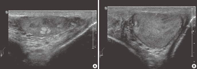
Ultrasonography of the left testis. (A) A 0.4 × 0.5 × 0.4 cm sized poorly defined, heterogeneous mass with increased echogenicity. (B) Hypoechoic ring identified around the mass forming a "tubule-in-a-tubule" appearance.

Ultrasonography of the left testis. (A) A 0.4 × 0.5 × 0.4 cm sized poorly defined, heterogeneous mass with increased echogenicity. (B) Hypoechoic ring identified around the mass forming a "tubule-in-a-tubule" appearance.