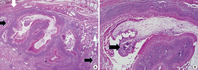
Fig. 2.

Histopathological findings of the left testis. (A) A sparganum surrounded by abscess and necrotic materials is present within the testicular parenchyma. White arrows indicate seminiferous tubules, a black arrow indicates tunica albuginea, and a white triangle rete testis (×12.5). (B) The sparganum shows tortous recess of the bothrium, tegument, longitudinal layer of the smooth muscle, and loose internal stroma and multiple calcarous bodies are present in the loose internal stroma (×40).
