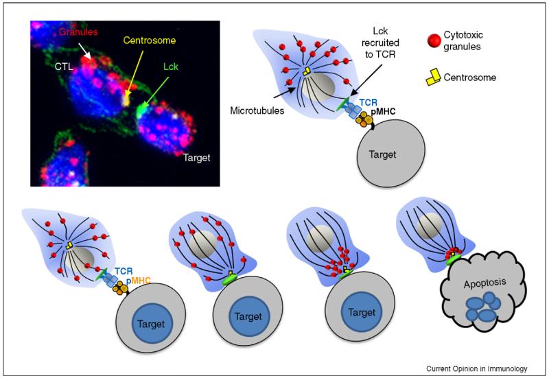
Figure 2. Mechanism of lytic granule release.
Recognition of the target MHC class I molecules bound to peptide antigen occurs via the TCR. Once the TCR is engaged, the most proximal signalling molecule to be recruited is Lck (shown in green). The centrosome (γ-tubulin, yellow), which is normally perinuclear, polarises towards the synapse and docks at the plasma membrane. The granules (LAMP-1, red) migrate towards the centrosome, where they dock and fuse with at the plasma membrane, releasing their contents into the synaptic cleft, causing target cell destruction.

