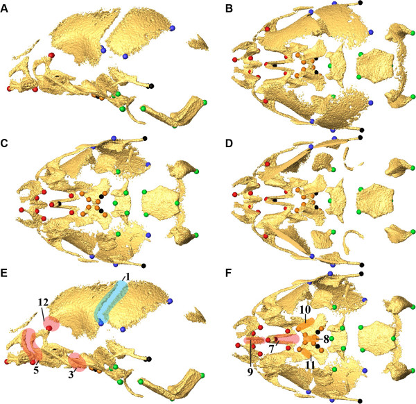
Figure 1.
Landmark and suture placement. Placement of anatomical landmarks (A-D) and sutures (E,F) on E17.5 mouse skull. Views are left lateral (A), superior (B), inferior (C), endocranial (D), left lateral (E), and inferior (F). Landmarks (A-D) are color coded by region: Face (red); Base (green); Vault (blue); Palate (orange). Additional landmarks that were used only in analysis of the global skull are shown in black. Sutures (E, F) are color-coded by region [Face (red); Vault (blue); Palate (orange)] and indicated by number: 1,2) Left, right coronal ; 3, 4) Left, right zygomatic-maxillary; 5,6) Left, right premaxillary-maxillary; 7) Intermaxillary; 8) Interpalatine; 9) Inter Premaxillary; 10, 11) Left, right maxillary-palatine; 12, 13) Left, right fronto-maxillary. Sutures 1-6 and 12-13 are bilateral; only left sutures are shown on the left lateral view (E). An additional table provides more complete definitions of the landmarks, as well as identification of the skull region in which each landmark is located [see Additional file 1]. More information on landmark identification and location can be found at: http://getahead.psu.edu/landmarks_new.html.

