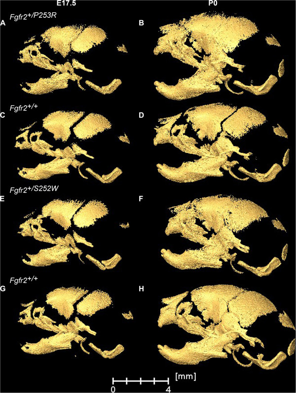
Figure 2.
Skull morphology of Fgfr2+/P253R and Fgfr2+/S252W Apert syndrome mice and unaffected littermates at E17.5 and P0. Left lateral views of 3D HRμCT reconstructions of representative mice from our study samples: Fgfr2+/P253R mutant at E17.5 (A) and P0 (B): unaffected littermate of the P253R model at E17.5 (C) and P0 (D); Fgfr2+/S252W mutant at E17.5 (E) and P0 (F); unaffected littermate of the S252W model at E17.5 (G) and P0 (H). By P0, midfacial retrusion is more severe and fusion of multiple sutures is apparent.

