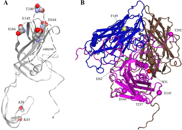
Figure 2.
Location of amino acids in the 3D structure of VP1 protein (A) and the complex (B) of VP1, VP2 and VP3. A and B are based on the PDB 3VBS which belonged to genotype C. the structure of PDB 3VBS was reconstituted with software of Cn3D (version 4.3.1).The consensus residues of K43, A58, S184 and T240 and canyon structure are shown in A; the variation sites of N31, E145, D164, I262 and T292 of VP1, F149 of VP2 are shown in B.

