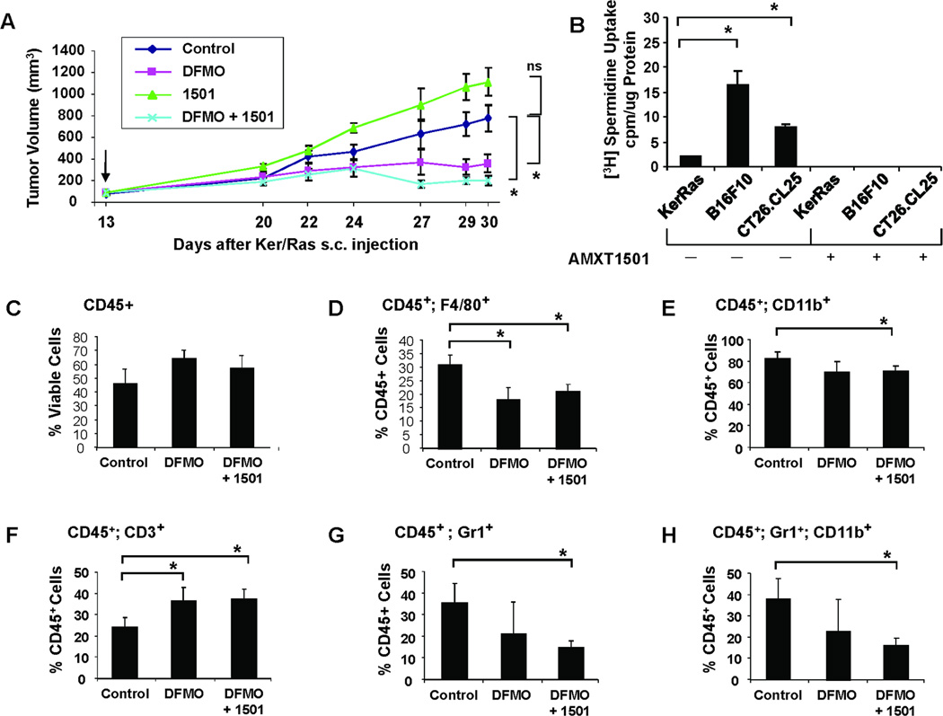
Fig. 4. PBT retards Ker/Ras tumor growth and stimulates macrophage infiltration in Ker/Ras tumors.
Primary murine keratinocytes (3 × 10⁶) transduced with v-H-Ras retrovirus were s.c. injected into syngeneic FVB mice. When the tumors were 50–100 mm3 in size, treatment was initiated with either saline, 0.5% DFMO (w/v) in the drinking water, AMXT1501 (i.p., 3 mg/kg, bid Mon-Fri and once a day on weekends), or co-treatment with DFMO and AMXT1501. A) Graph shows tumor growth rate and volume in mice bearing Ker/Ras tumors under different treatments (n = 5 mice/treatment group; mean volume per time point ± SEM). *, P < 0.01. B) To compare basal polyamine transport in cultures of Ker/Ras cells with B16F10 and CT26.CL25 cells, 3H-spermidine was added, and cells were incubated for 1 hr at 37°C or on ice. Uptake of 3H-spermidine (cpm) was measured using a scintillation counter and is normalized to protein. Values are the mean of triplicate samples ± SD. *, P < 0.01. (C–H) Flow cytometry analyses of CD45+ inflammatory infiltrates in Ker/Ras tumors. Mice with Ker/Ras tumors were treated for two weeks with DFMO or with both DFMO and AMXT1501, and tumors were analyzed by flow cytometry for C) % CD45+ immune cell infiltrates in total viable tumor cell population. Subpopulations of viable CD45+ cells were also stained for the following immune cell markers: D) F4/80+ macrophages; E) CD11b+ myeloid cells; F) CD3+ lymphocytes; G) Gr-1+ granulocytes; and H) Gr-1+CD11b+ MDSCs. *, P < 0.01.

