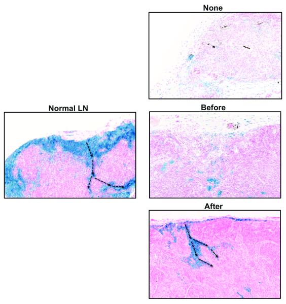
Figure 3. Sterile inflammation after lymph node transfer improves lymphatic function.
Representative histological images (20x magnification) of ferritin staining in lymph nodes harvested from animals in various experimental groups (blue stain). Note subcapsular pattern of staining and drainage into medullary area in normal lymph node and animals in which inflammation was induced after lymph node transfer (black arrows show direction of flow). Note relative lack of staining in lymph nodes without inflammation or in those in which inflammation was induced prior to tissue transfer.

