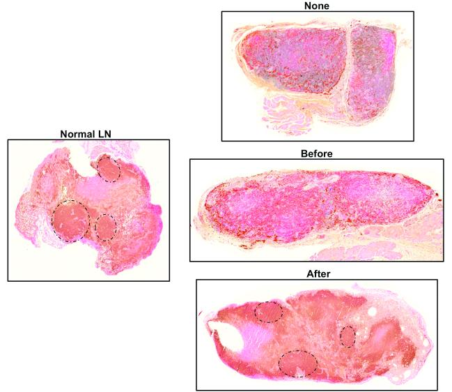
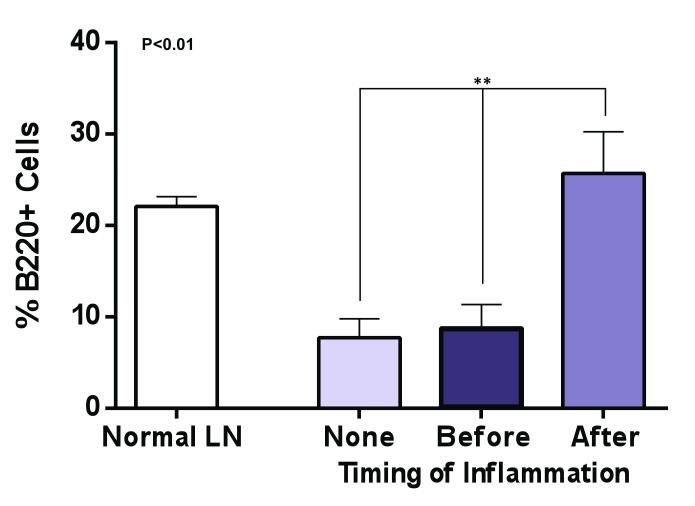
Figure 5. Inflammation after lymph node transfer preserves B cell follicular architecture.
A. Representative photomicrographs (5x magnification) of lymph nodes from various groups stained for B220, a B cell marker. Note follicular pattern of B cell clusters in normal lymph node and in lymph node harvested from animals in which inflammation was induced after transfer (black circles).
B. Quantification of B cells (percentage of B cells as a function of lymph node area). Note significant increase in B cells in inflammation after transfer group as compared with no inflammation or inflammation before transfer groups.


