
Figure 1.
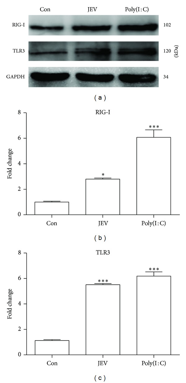
Expression of TLR3 and RIG-I in JEV-infected BV-2 cells. (a) BV-2 cells were either mock infected or infected with JEV at an MOI of 1. Poly(I : C) was added as a positive control. Western blotting was performed to detect TLR3 and RIG-I at 24 hpi. (b, c) The protein levels were quantified with immunoblot scanning and normalized to the amount of GAPDH. Error bars represent the standard deviation of results from three independent assays (*P < 0.05; ***P < 0.001).