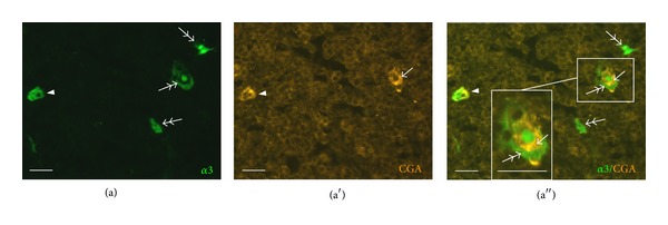
Figure 6.

Immunolabeling for CGA. Both double- (eGFP+/CGA+, arrowhead) and single-labeled cells (eGFP+/CGA−, double-headed arrow; eGFP−/CGA+, arrow) can be observed. CGA-immunoreactive and eGFP-positive cells occur together in Hassall's corpuscle-like structures (higher magnification in inset). Scale bar, 20 μm.
