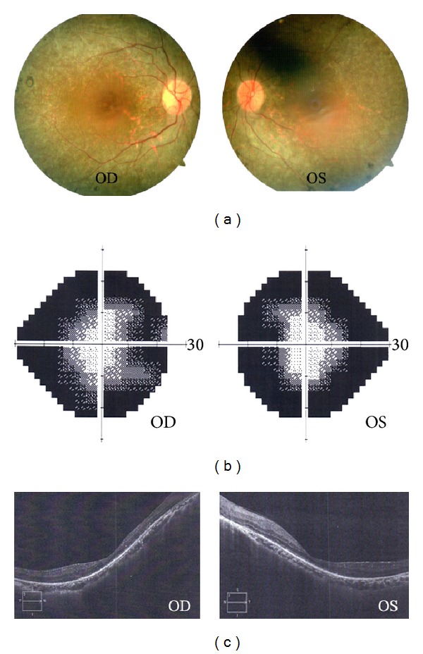
Figure 2.

Representative clinical characteristics of the proband. (a) Fundus photographs show bone spicule-like pigmentation and retinal vascular attenuation. (b) Visual field test point locations show the loss of peripheral visual field. (c) Optical coherence tomographic (OCT) images show severe thinning of the photoreceptor inner/outer segment.
