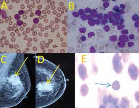
Figure 1. Peripheral blood smear and bone marrow aspirate(A&B) show lymphoblasts with coarse chromatin and highnuclear cytoplasmic ratio (Jenner-Giemsa stain, 1000x).Mediolateral oblique and cranio-caudal views of themammogram show homogeneous mass with architecturaldistortion and microcalcification (C&D). Fine-needleaspiration shows immature lymphoid cell with duct (400X)(E).

