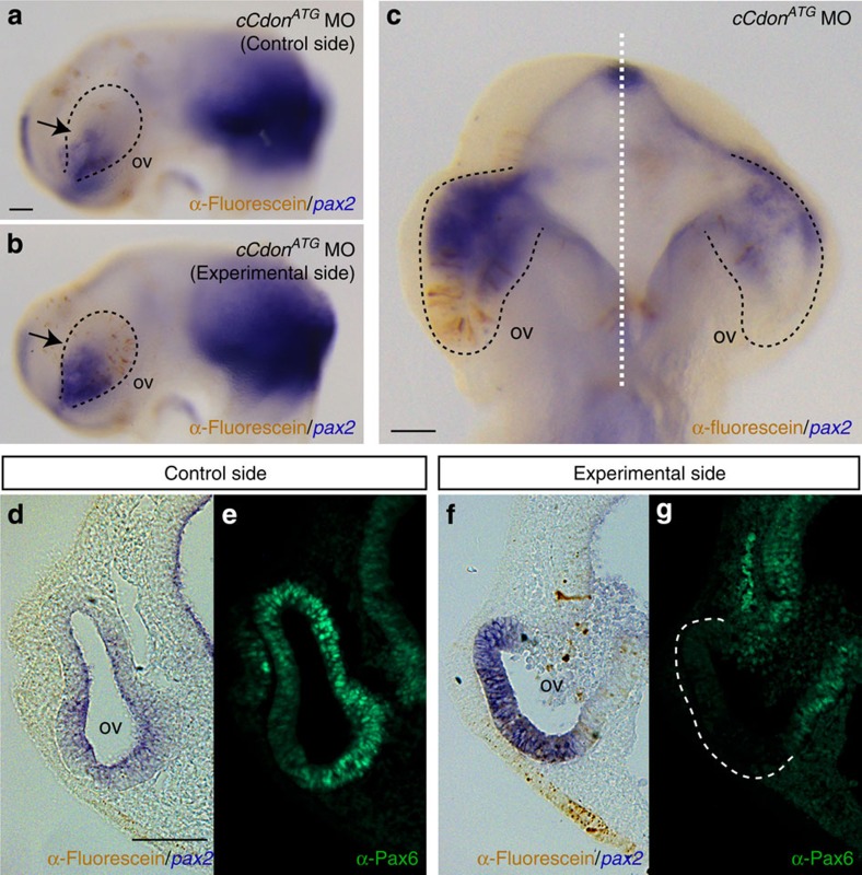
Figure 5. Localized interference with Cdon expression in the optic vesicles expands the distal optic stalk domain.
(a–f) Lateral (a,b) and ventral (c) views of the control (a) and experimental (b) optic vesicles (ov) of HH14 chick embryos with unilateral focal electroporation of a carboxyfluorescein conjugated cCdonATG MO at HH8. Embryos were hybridized for pax2 (blue signal) and immunostained with anti-fluorescein antibodies (brown signal) to detect MO distribution (a–d,f). Pax6 immunohistochemistry was performed in cryostat sections of electroporated embryos (e,g). Pax2 expression is expanded to the entire optic vesicle of cCdonATG MO-treated embryos (b,c) in comparison with the non-electroporated control eye (a,c). Pax2 expansion is associated with a reduction of Pax6 distribution in the retina (f,g) in contrast to a wild-type condition (d,e). The optic vesicles are outlined with black (a–c) or white (g) dashed lines. Scale bar, 50 μm.

