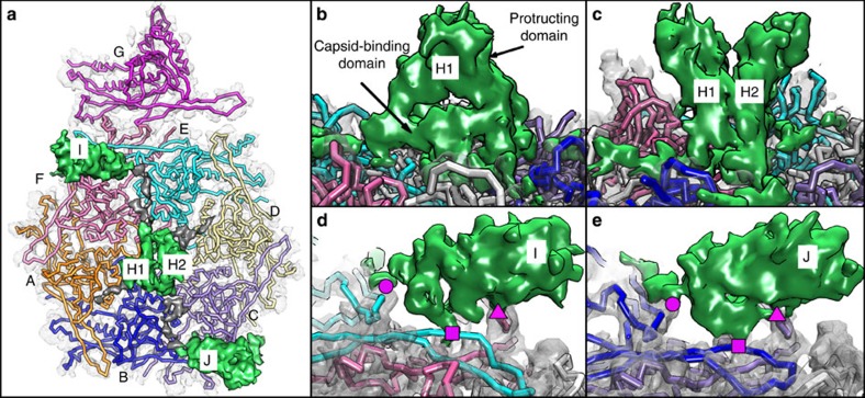
Figure 4. Binding sites of I/H/J.
(a) One asymmetric unit of Syn5 consisting of 11 polypeptide chains, labelled as A–J, where the two-polypeptide chains of H are labelled as H1/H2. The seven major capsid protein gp39 models (A–G) follow the colour scheme of Fig. 2b. Here the knob-like proteins (green) are seen connected by grey densities. (b) The binding site of the density H to gp39 (a side view such that only one monomer H1 is seen). (c) A close up of H in a 90° rotated view of (b) (a side view such that both monomers H1/H2 are seen). (d,e) A close up of the equivalent binding sites of densities I and J to gp39, annotated by magenta circle, square and rectangle symbols.

