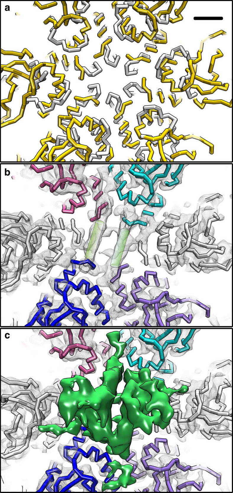
Figure 7. Gp58 (H1/H2) incorporation.
(a) Difference between the opening at the centre of the hexamer in Syn5 (gold) and that seen in HK97 (white). Scale bar, 13Å. (b) The same region of (a) in Syn5 map (slice view, grey mesh) with fitted Syn5 model (white). Here two helical densities (green cylinders) are observed connecting four gp39 subunits (coloured). (c) A non-slice view of (b) to illustrate that the helical densities in (b) correspond to the capsid-binding domain of gp58 (green) contributed by its H1 and H2 monomers.

