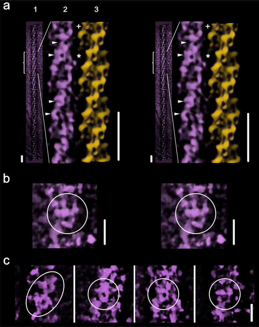
Figure 6.
Wide-eye stereo views of (a): (1) the near outer shell of the tomographic 3D-reconstruction of one thick filament; (2) an enlarged view of (1), filtered and cylindrically masked (box, Fig. 1b); and (3) an equivalent segment of the SF-IHRSR 3D-reconstruction calculated from the tomogram images subset (Fig. 4a). The asterisk points the MIH motif used to fit the 3DTP model into the tomographic 3D-reconstruction shown in Fig. 5b and Supplementary movie 2. Bars 43.5 nm. The near outer shell of the tomogram reveals in some places shapes (labeled with 4 arrowheads and a “+”) which approximately resemble the MIH motif. The one labeled with a “+” is best seen after left hand rotation of the region by ~ 21° as shown in the enlarger wide-eye stereo view (b) (see Suppl. movie 3). A panel of several shapes (labeled with arrowheads Fig. 6a) is shown in (c). Bars 14.5 nm for (b) and (c)

