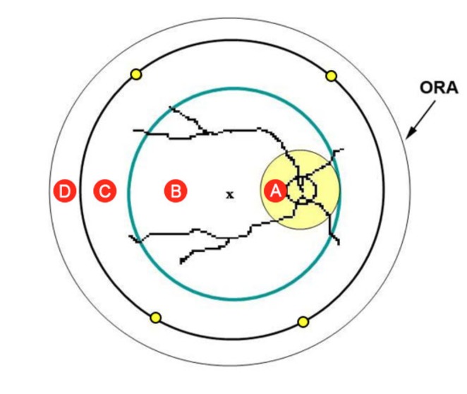
FIGURE 1.
Schematic of retinal hemorrhage assessment tool. Retina is divided into four zones: A (yellow circle), B (green circle), C (black circle), and D (peripheral retina beyond zone C). The nasal edge of zone B is tangential to the nasal edge of zone A. The edges of the midperipheral zone C run approximately through the vortex veins (yellow dots). The x represents the fovea.

