. 2014 Jun 23;3:e02687. doi: 10.7554/eLife.02687

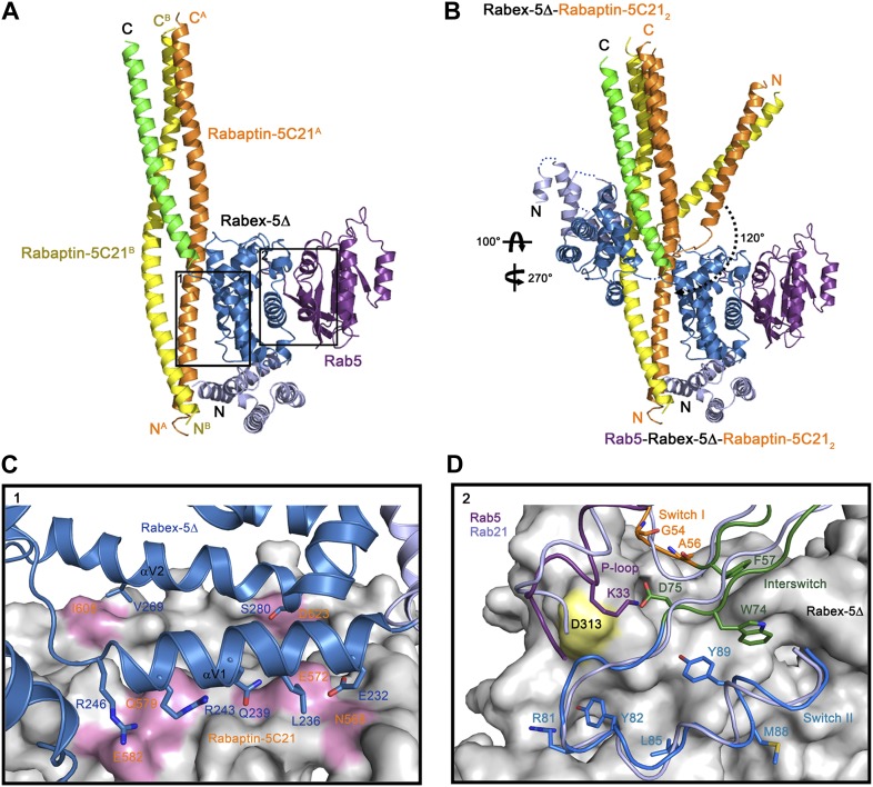
Figure 2. Crystal structure of the Rab5-Rabex-5Δ-Rabaptin-5C212 complex.

(A) A ribbon representation of the overall structure of the Rab5-Rabex-5Δ-Rabaptin-5C212 complex. Only one Rab5-Rabex5Δ-Rabaptin5C212 complex in the asymmetric unit is shown with Rab5 in purple and Rabex-5 and Rabaptin-5C21 in the same colors as in Figure 1A. (B) Comparison of the Rab5-Rabex-5Δ-Rabaptin-5C212 complex and the Rabex-5Δ-Rabaptin-5C212 complex based on superposition of the three-helix bundle formed by Rabex-5CC and the C-terminal regions of Rabaptin-5C212. (C) Interactions between the Rabex-5 GEF domain and Rabaptin-5C212. The GEF domain is shown in ribbon representation in blue and the interacting residues are shown with side chains. Rabaptin-5C212 is shown in surface representation with the interacting residues colored in pink. (D) Interactions between Rab5 and the Rabex-5 GEF domain. Rab5 is shown in coil representation with the P-loop, switch I, switch II, and interswitch region colored in purple, orange, blue, and dark green, respectively. Several key residues are shown with side chains. The GEF domain is shown in surface representation with Asp313 colored in yellow. For comparison, Rab21 in its complex with the Rabex-5 GEF domain (Delprato and Lambright, 2007) is shown in coil representation in light blue.

DOI: http://dx.doi.org/10.7554/eLife.02687.012

Figure 2.

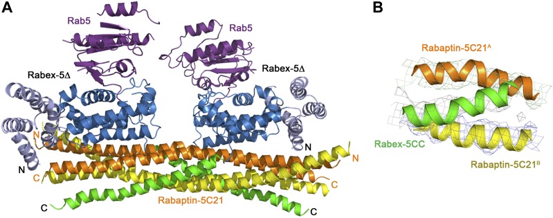
Figure 2—figure supplement 1. Crystal structure of the Rab5-Rabex-5Δ-Rabaptin-5C212 complex.

Figure 2—figure supplement 1.

(A) A ribbon representation of the overall structure of the dimeric Rab5-Rabex-5Δ-Rabaptin-5C212 complex in an asymmetric unit. There are two Rab5-Rabex5-Rabaptin5C212 complexes related by a two-fold non-crystallographic symmetry in the asymmetric unit. Rab5, Rabex-5Δ, and Rabaptin-5C21 are colored the same as in Figure 2A. Rabaptin-5C21 and Rabex-5 are well defined in the electron density map; however, only one Rab5 is fairly defined while the other is poorly defined. (B) Representative simulated annealing composite omit map of the Rab5-Rabex-5Δ-Rabaptin-5C212 complex. The map is contoured at 1.0σ with the final structure shown in ribbon model.
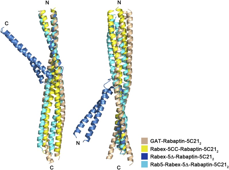
Figure 2—figure supplement 2. Superposition of Rabaptin-5C212 in different complexes.

Figure 2—figure supplement 2.

Rabex-5CC-Rabaptin-5C212: yellow, Rabex-5Δ-Rabaptin-5C212: blue, Rab5-Rabex-5Δ-Rabaptin-5C212: cyan, and GAT-Rabaptin-5C212 (PDB code 1X79) (Zhu et al., 2004a): light orange. The N-terminal regions of Rabaptin-5C212 in these complexes can be superimposed well (RMSD of ∼1.0 Å for 31 Cα atoms) (left panel); however, the C-terminal regions cannot (RMSD of ∼3.6 Å for 42 Cα atoms) (right panel). The angle between the two regions also differs substantially which is about 40° in the Rabex-5Δ-Rabaptin-5C212 complex, and about 180° in the Rabex-5CC-Rabaptin-5C212 complex, the Rab5-Rabex-5Δ-Rabaptin-5C212 complex, and the GAT-Rabaptin-5C212 complex.