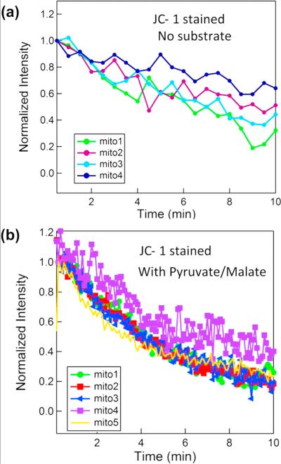
Figure 6.
Fluorescence intensity measurement of JC-1 stained mitochondria. a) Substrates are not used. b) OXPHOS substrates (5 mM pyruvate and 5 mM malate) are added to respiration buffer just before flowing the mitochondria into the nanofluidic channel. This activates the electron transport chain and increases the mitochondrial membrane potential Δψm initially.

