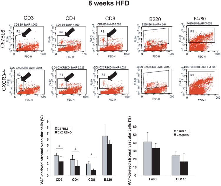
Figure 3.
Peri-epididymal adipose tissue from obese CXCR3-deficient mice contains fewer T cells than obese controls after 8 weeks of high-fat diet, as detected by flow cytometry.
Stromal vascular cells from 1 g of adipose tissue of CXCR3-deficient mice and wild-type control mice fed a high-fat diet (HFD) for 8 weeks were labeled with conjugated antibodies to CD3, CD4, CD8, B220, F480, and CD11c, and analyzed by flow cytometry. The graphs represent percentages of gated cells in the CXCR3-deficient group (black bars) and the control group (gray bars). Data are shown as mean ± SD. *p <0.02; n=9/group.

