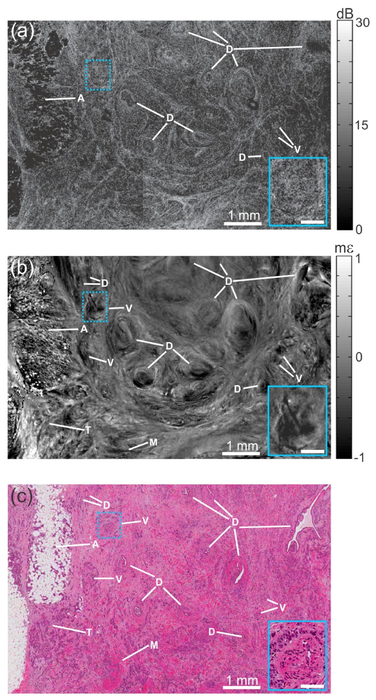
Fig. 6.

Optical coherence micro-elastography of malignant human breast tissue. (a) En face OCT image at a depth of ~100 μm. (b) Corresponding en face micro-elastogram. (c) Histology, co-registered with OCT and micro-elastogram. A, adipose; D, duct; M, smooth muscle; T, region densely permeated with tumor; and V, blood vessel. In the micro-elastogram, the scale is in millistrain, mε. The insets show a 2.5× magnification of the blue-dotted boxes. Scale bars in the inset, 0.25 mm. In the micro-elastogram, the scale is in millistrain, mε.
