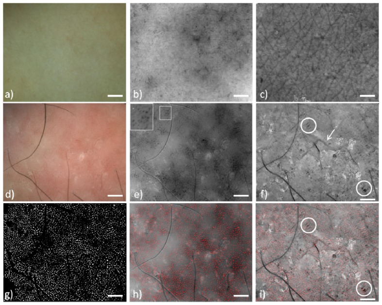
Fig. 2.
Polarization Multispectral Dermoscope images. Healthy skin (a-c). Psoriasis (d-i). a)Dermoscopic white light image, b) Blood Contrast, blood vessels are encoded dark grey, small ones are visible c) Scattering Contrast, well defined wrinkle network is visible, d) Dermoscopic white light image where homogeneous red dots with some patches of pink to bright red are visible, e) Blood Contrast image, bigger blood vessels are visible. Inset: magnified vessel details, f) Scattering Contrast image. The wrinkle network is completely absent. Arrow: wrinkle, circles: dark parallel structures g) The vessel segmentation image, f) Blood Contrast image merged with the segmented image, i) Scattering Contrast image merged with the segmented image, blood vessels do not colocalize with the characteristic dark parallel structures of psoriasis. (Scale bar: 1mm)

