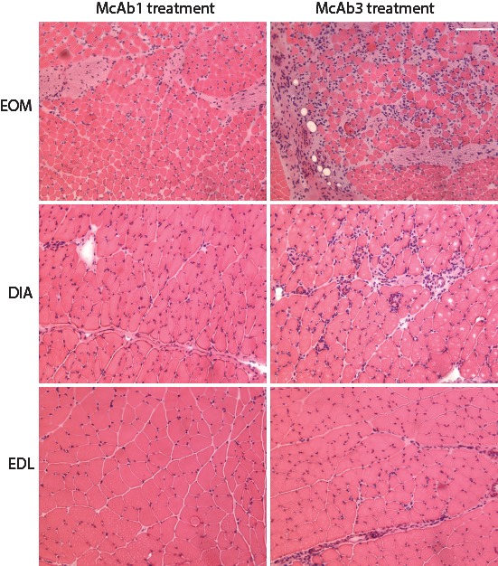
Figure 4.

Histological sections stained with hematoxylin and eosin and viewed at low magnification to provide a representative view from EOM, EDL, and DIA from control and McAb-3 injected mice. Note more extensive inflammatory inflammation in EOM compared with other muscles.
