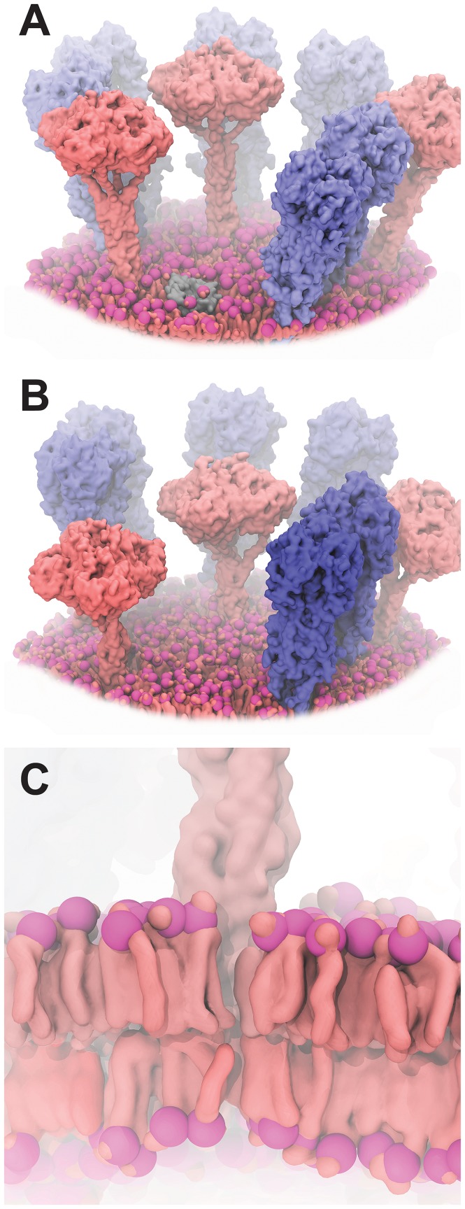
Figure 5. Models of influenza surface patches.
NA and HA glycoproteins are shown in pink and blue, respectively. Large-scale curved bilayers were modeled using LipidWrapper. To improve clarity, some portions of the bilayers have been removed, and colors have been enhanced. A) A model including wild-type (long-stalk) NA. B) A model including a shorter NA, caused by a twenty-amino-acid deletion in the NA stalk. C) A close-up stylized view of the atomistic LipidWrapper-generated bilayer.

