
Fig. 2. Non-denaturing electrophoresis stained for hydrolytic activity of pooled microsomes from human (A), mouse (B) or rat (C) livers.
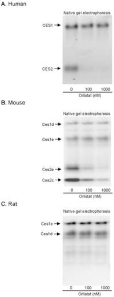
Microsomes (15 μg) from human liver (n = 14), mouse liver (n = 4) or rat liver (n = 4) were pre-incubated in a total volume of 10 μl with orlistat at various concentrations (0, 0.1 or 1μM) for 30 min. To the pre-incubated mix, 2.5 μl sample loading buffer was added. The samples were then subjected to native gel electrophoresis and stained for esterase activity with 1-naphythylacetate as described in the section of Materials and Methods. The staining intensity was captured by Carestream 2200 PRO Imager. This experiment was repeated four times.