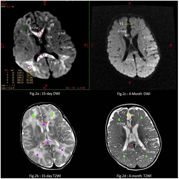
Figure 2. MRI scans of a four-month child with CHIKV neonatal encephalopathy.
- Child n°4 of Table 5. Full-term small for gestational age 38-week neonate. m5-Apgar score: 10/10. Encephalopathy with sepsis and DIC on day 4. Global developmental delay with DQ = 77 and microcephaly (head circumference 43 cm, −1.5 z-score SD) at 20 months. Axial sections via the interventricular foramen at day 15 on the left side: Diffusion-weighted imaging (DWI) with apparent diffusion coefficient (ADC) map (Fig. 2a), and T2-weighted imaging (T2WI) (Fig. 2b). Axial sections via the body of third ventricular at month 4 on the right side: DWI with ADC map (Fig. 2c), and T2-weighted imaging (Fig. 2d). MRI scans show scattered areas of cytotoxic edema (violet circles) with decreased-diffusion signals on the ADC map or normal-appearing white matter (green circles) at 15-day scans, absence of persistent brain swelling (normal ADC) but scattered demyelination with scalloped-appearance of white matter atrophy (green triangles) including thinning of the corpus callosum (double red lines), passive dilatation of supratentorial interhemispheric subarachnoïd spaces (double yellow arrows). Anatomic abbreviations: WM: white matter; frontal lobes: superior frontal (F1), cingular (CingG), inferior frontal (F3), post-central (PoC), Th: thalamus, hCN: head of the caudate nucleus; parietal lobes: supra marginalis (SuMa), angular (Ang), superior parietal (P1) gyri; genu (gCC) and splenium (sCC) of the corpus callosum; occipital lobe (Cuneus); aLV: atrium of the lateral ventricule containing the choroid plexuses.

