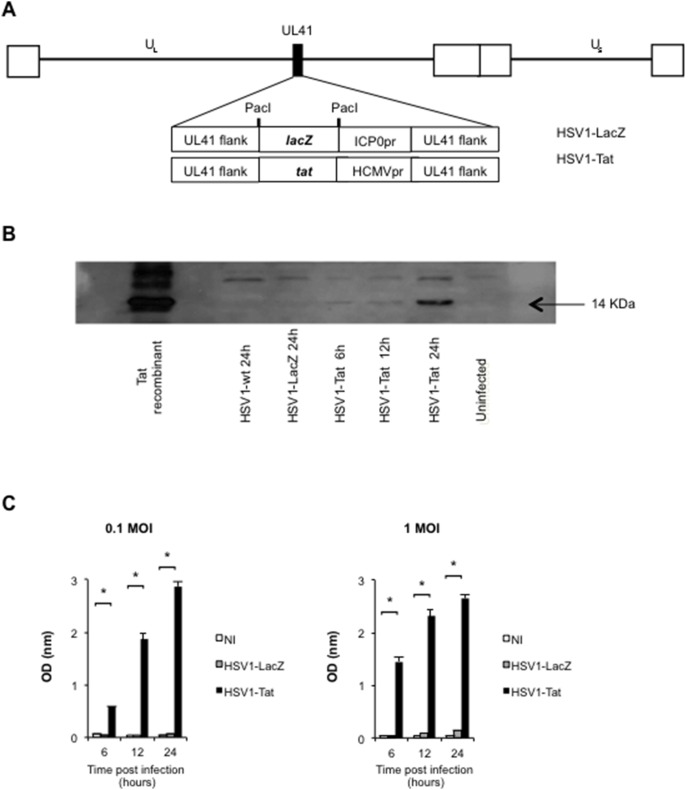
Figure 1. Vector representation and analysis of biological Tat expression.
Schematic representation of the HSV1-LacZ/HSV1-Tat recombinant viruses (A). The black square in the HSV1 backbone indicates the UL41 locus (vhs gene), which has been deleted by insertion of the lacZ- or Tat-promoter cassette, respectively. The white squares represent the terminal and internal repeats of the HSV genome delimiting the unique regions (UL: unique long; US: unique short). Analysis of Tat expression by HSV1-Tat recombinant vector (B). Western blot analysis of BALB/c cells infected with HSV1-Tat (1 MOI) after 6, 12 and 24 h post-infection. Negative controls are represented by uninfected cells and by cells infected with wild-type HSV1 or HSV1-LacZ. Recombinant Tat protein (Tat) was used as positive control. Analysis of biological Tat activity by CAT ELISA assay (C). CAT expression induced by functional Tat protein produced by the HSV1-Tat viral vector was measured 6, 12 and 24 h post-infection in HeLa3T1 cell extracts infected with HSV1-Tat (0.1 or 1 MOI). Negative controls were uninfected cells and cells infected with wild-type HSV1-LacZ. The means (± SD) of six independent experiments are shown.

